Microscopes & Imaging
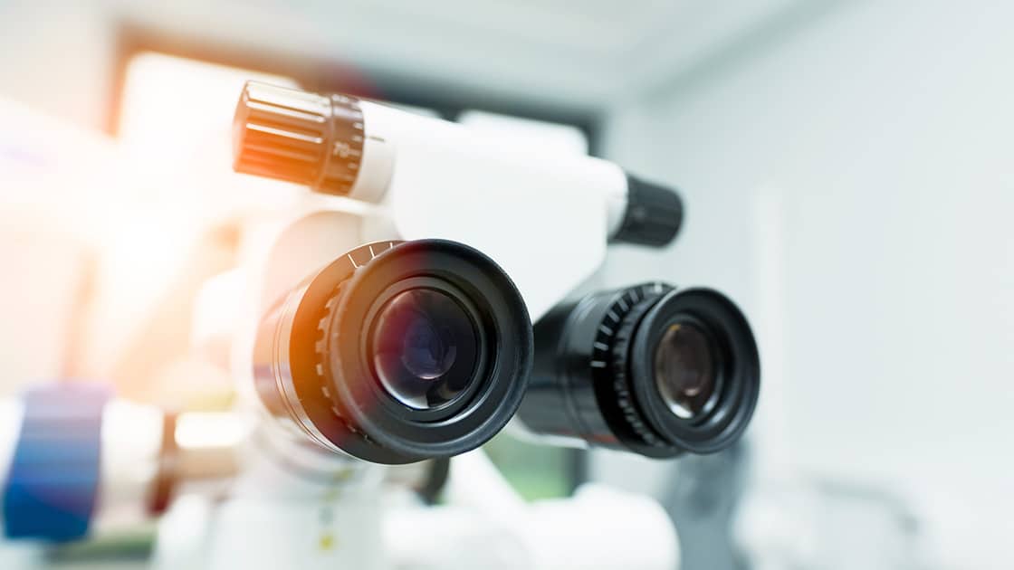
The use of specialized operating microscopes means that the doctor is able to get a detailed look at the work they are doing during all phases of your endodontic treatment. The additional magnification and illumination allow them to work with great precision and see small details such as calcified canals and fractures. The Endodontist is able to more accurately diagnose and treat the patient using a dental surgical microscope to improve the potential outcome of the treatment from “good” to “excellent”. Further, some microscopes may be equipped with high resolution video and digital photography allowing the doctor to enhance patient communication and document treatment.
Electronic Apex Locator
This device is used to obtain the proper length of the root canal, from which the doctor can determine the position of the file relative to the apex of the root. This can help ensure that the canal is completely free of debris, reducing potential future complications.
Cone Beam Scanner 3D Imaging
3D imaging provides better quality and more detailed images than traditional x-rays, which leads to improved diagnostic and treatment planning abilities. This technology is also less invasive and emits less radiation than traditional x-ray machines.
Digital Flat Screen Monitors
These monitors are found next to every patient chair. Patients can watch a movie or TV show. Patients can also view their dental radiographs when speaking to the doctor about the findings for a better understanding of their oral health.
Looking for an endodontist in the Oregon City, Gladstone, Clackamas, Happy Valley, Milwaukie, Canby, West Linn, Lake Oswego, Wilsonville, Molalla, or Estacada areas? Contact us at
to schedule an appointment today!
